
Introduction
Erythema Dyschromicum Perstans (EDP), also known as ashy dermatosis, is an idiopathic, painless pigmentary skin condition of dermal origin.1 In the literature, there have only been a few reports of unilaterally distributed EDP especially those with a well-demarcated eruption in a localized region.2 Although cases are reported worldwide, the disorder is most prevalent in South America and South-Central USA, particularly in Hispanic populations and individuals with Fitzpatrick Skin type III-IV.2–4 It presents with gray macules and patches of varying shape and size that are sometimes surrounded by a slightly raised erythematous border. In nearly all cases of this disease, it is distributed symmetrically, often on the face, neck, trunk, and arms.5,6
Case Synopsis
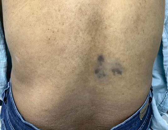
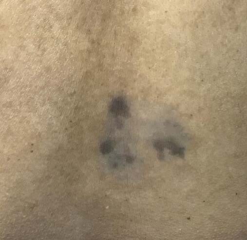
A 47-year-old Hispanic woman presented to our clinic with concerns regarding the appearance of a dark rash on her lower back that had been progressing for the past two months. She denied associated itching or pain and had not experienced anything similar previously. In addition, she had no history of injury to the area and had applied nothing topical. Upon examination, dark gray-blue macules were evident and superimposed upon a larger, light gray patch, localized to the patient’s right lower back (Figure 1-2).
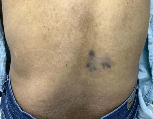
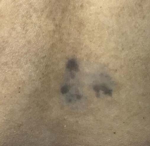
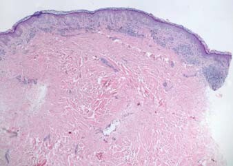
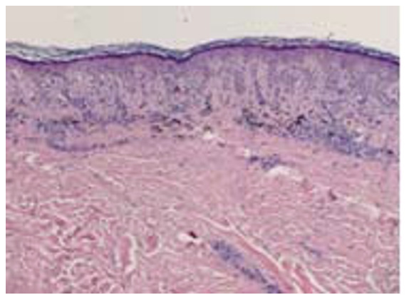
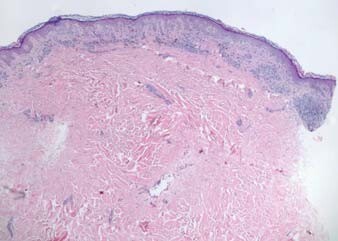
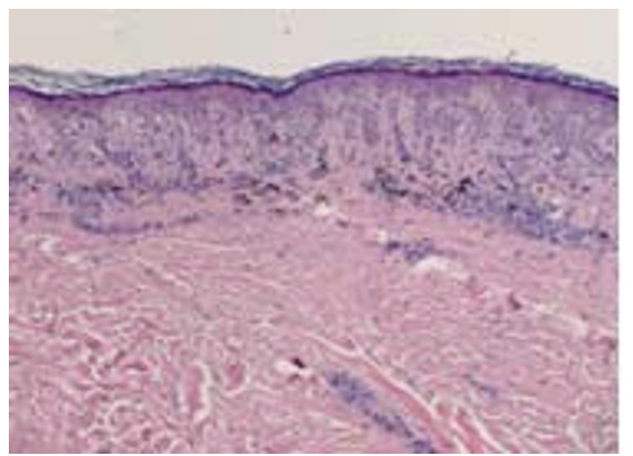
Following this clinical presentation, the list of possible differential diagnoses included lichen planus pigmentosus, post-inflammatory hyperpigmentation (PIH), erythema dyschromicum perstans, lichen planus, and macular amyloidosis. To elucidate an accurate diagnosis, a 4 mm punch biopsy was performed on one of the hyper pigmented macules. Histology ruled out most differential diagnoses aside from EDP with the possibility of PIH included; however, PIH was clinically excluded by the patient’s denial of past inflammation or injury to the site (Figure 3-4).
The patient is currently being treated with cycles of triamcinolone acetonide 0.1% cream applied twice a day to the area for two weeks at a time followed by a one-week break, during which time pimecrolimus 1% cream is applied to the area twice per day. Her progress has not yet been evaluated in follow-up.
Discussion
EDP is a rare, slowly advancing skin condition that has mostly been diagnosed in women of Hispanic descent, matching the demographics of the presented patient.7 In addition to the clinical presentation, histopathological evaluation of the disease is helpful when confirming the diagnosis of EDP.8 Featured histological findings seen in patients with EDP include vacuolization of the basement membrane zone, perivascular lymphocytic infiltration, papillary dermal edema, dermal melanophages, necrotic keratinocytes in the basal layer, and a focal or lichenoid pattern with colloid bodies along the dermo epidermal junction.5,9,10 The patient’s biopsy specimen demonstrated a mild lichenoid pattern with papillary dermal macrophages and aligned with classic histopathological findings of EDP. While her demographics and histology coincided well with the prototypical presentation of EDP, our patient’s uniquely unilateral presentation is noteworthy.
Conclusion
This case seeks to portray a distinctive way that erythema dyschromicum perstans can present that has not been commonly considered in the literature previously. The cause of unilaterally distributed EDP is not well understood. Nevertheless, practitioners should include EDP in the differential diagnosis for patients with symmetric, as well as unilateral pigmentary changes.
Conflicts of interest disclosure
The primary and co-authors declare no conflicts of interest and no financial support received through his research activity.
Ethical statement
Verbal and written consent for writing case reports and publication was taken from patient.