
Introduction
Cerebrovascular stroke is the leading global cause of long-term motor impairment in adults and represents a major source of disability adjusted life years worldwide.1 While both children and adults experience neurological injury following stroke, the superior compensatory capacity of the pediatric nervous system is well established, underscoring the importance of understanding the mechanisms and limits of neuroplasticity in adult patients.2,3
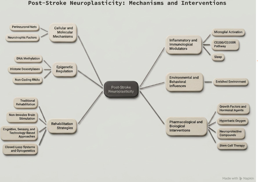
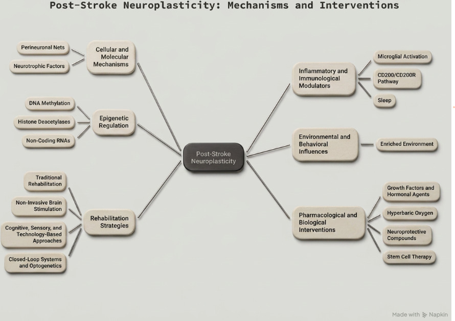
Neuroplasticity broadly defined as the ability of the nervous system to reorganize its structure, function, and connections in response to intrinsic or extrinsic stimuli, which is the cornerstone of post-stroke recovery. In adults, this capacity is modulated by a complex interplay of molecular, cellular, epigenetic, and environmental factors. Understanding these mechanisms is essential not only for elucidating the biology of recovery but also for developing targeted therapeutic interventions. This review systematically examines the current body of literature on adult post-stroke neuroplasticity, organized around five domains: (1) cellular and molecular mechanisms, (2) inflammatory and immunological modulators, (3) epigenetic regulation, (4) environmental and behavioral influences, and (5) therapeutic strategies encompassing rehabilitation, pharmacology, and emerging technologies.
1. Cellular and Molecular Mechanisms of Post-Stroke Neuroplasticity
1.1. Perineuronal Nets and Inhibitory Extracellular Matrix Remodeling
Following a vascular insult, there is a differential expression of inhibitory molecules, most notably Chondroitin Sulfate Proteoglycans (CSPGs), in the peri-infarct region. These molecules are organized into structures known as Perineuronal Nets (PNNs) extracellular matrix complexes that serve a stabilizing role in the mature nervous system but concurrently inhibit neuronal plasticity after injury.4
Recovery at the cellular level involves the action of proteases on these inhibitory structures. Tissue plasminogen activator (tPA) acts on ADAMTS-4 (A Disintegrin and Metalloproteinase with Thrombospondin motif type 4), a member of a proteinase family capable of degrading inhibitory CSPGs. Both ADAMTS-4 and select members of the Matrix Metalloproteinase (MMP) family contribute to extracellular matrix reorganization and thereby facilitate neuronal recovery.4
1.2. Neurotrophic Factors and Signaling Pathways
Brain-Derived Neurotrophic Factor (BDNF) is among the most extensively studied neurotrophic factors in post-stroke recovery. Following elaboration by neurons, BDNF binds to membrane lipid rafts (MLRs) discrete microdomains enriched in the structural protein Caveolin. This interaction initiates downstream activation of multiple signaling cascades, including PI3K/Akt, MAPK/ERK, NF-κB, Sonic Hedgehog, cAMP, Notch, and Wnt/β-Catenin pathways, all of which contribute to synaptic plasticity, neuronal survival, and axonal remodeling.5
2. Inflammatory and Immunological Modulators
2.1. Microglial Activation and Post-Stroke Inflammation
Microglial activation following ischemic injury generates a pro-inflammatory milieu that has been documented to impair neuroplasticity. Multiple studies have reported improved functional outcomes following administration of anti-inflammatory agents, supporting the notion that post-stroke neuro-inflammation constitutes a significant barrier to recovery.6
2.2. The CD200/CD200R Pathway
The CD200/CD200R signaling axis, known to suppress microglial activation and reduce elaboration of pro-inflammatory cytokines, has been implicated in facilitating spontaneous functional recovery after stroke.7 This pathway represents a potential immunomodulatory target for augmenting the neuroplastic response in the post-acute phase.
2.3. Sleep, Inflammation, and Neuroplasticity
Sleep disorders are associated with elevated levels of inflammatory and oxidative stress biomarkers and have been documented to adversely affect stroke evolution and functional recovery.8,9 In contrast, slow-wave sleep has been positively correlated with recovery outcomes, with evidence that the restorative architecture of sleep actively promotes neuroplastic processes.10 These findings suggest that sleep optimization may be an underutilized adjunct in post-stroke rehabilitation.
3. Epigenetic Regulation of Neuroplasticity
3.1. Non-Coding RNAs and Emerging Molecular Targets
MicroRNA (miRNA) expression in neural tissues is spatially variable across the ischemic core and penumbra, with different miRNA subtypes exerting divergent effects on neuroprotection versus cell death.11 Beyond miRNAs, circular RNA HIPK2,12 the serotonin transporter gene SLC6A4,13 and C-C Chemokine Receptor 5 (CCR5),14 have recently been implicated in post-stroke neuroplasticity, opening novel molecular avenues for therapeutic targeting.
3.2. Histone Deacetylases
Histone deacetylase (HDAC) expression is altered following ischemic injury. Evidence from multiple studies indicates that inhibition of HDAC-2 promotes transcriptional activity in neural tissue and is associated with improved functional recovery post-stroke,15–17 these findings have stimulated interest in HDAC inhibitors as candidate neuroprotective and neurorestorative agents.
3.3. DNA Methylation
Global polymorphisms increase in DNA methylation have been observed in infarcted tissues following stroke. Hyper methylation of the BDNF promoter region, in particular, has been linked to attenuated neurotrophic signaling and poorer functional recovery establishing epigenetic silencing as a pathological mechanism in post-stroke neuroplasticity impairment.18,19
4. Environmental and Behavioral Influences
4.1. Enriched Environment
Enriched environment (EE), defined as an environmental modification designed to provide multi-sensory stimulation, and has been shown in preclinical models to promote Perineuronal net formation around parvalbumin containing GABAergic (PV/GABA) neurons in the neocortex.20 Additionally, EE modulates the proteolytic activity and mRNA expression of extracellular matrix proteases and their inhibitors in the somatosensory cortex.4 These findings provide a mechanistic basis for the efficacy of activity-dependent rehabilitation and identify candidate molecular targets for enhancing rehabilitative outcomes in human patients.21
5. Rehabilitation Strategies
5.1. Traditional Rehabilitation Modalities
Established rehabilitation approaches including speech therapy,22,23 physical therapy,1,23 occupational therapy,23 constraint induced movement therapy (CIMT) 23 and gait therapy24 remain the foundation of post-stroke recovery programs. These modalities capitalize on activity dependent plasticity and are most effective when initiated within the critical therapeutic window.
5.2. Non-Invasive Brain Stimulation
Non-invasive brain stimulation techniques offer a means to modulate cortical excitability and promote neuroplasticity beyond what conventional therapy achieves alone. A key mechanism underlying these benefits involves the attenuation of interhemispheric inhibition, wherein the contralateral (unaffected) hemisphere exerts inhibitory influence over the damaged hemisphere which tDCS and TMS can specifically target.25 Transcranial Direct Current Stimulation (tDCS) and Transcranial Magnetic Stimulation (TMS), including its repetitive form (rTMS) have demonstrated efficacy as adjuncts to traditional rehabilitation in multiple randomized trials.3,25–29
5.3. Cognitive, Sensory, and Technology-Based Approaches
Cognitive rehabilitation strategies including mental practice, mirror therapy, and motor imagery1 augment motor recovery by engaging neural circuits involved in movement planning and execution. Sensory stimulation modalities such as High-Frequency TENS, Low-Frequency TENS, Electro-acupuncture, and Neuromuscular Electrical Stimulation provide afferent input to promote cortical reorganization.25
Technology-based interventions, including augmented and virtual reality platforms,30,31 brain-computer interfaces (BCIs) and robotic-assisted therapy, enable high-intensity, repetitive, task-specific practice that can be tailored to individual deficits. Vagal nerve stimulation has also demonstrated efficacy as an adjunct neuro-modulatory approach.23,32,33
5.4. Closed-Loop Systems and Optogenetics
Emerging neurorehabilitation paradigms include closed-loop systems that provide real-time biofeedback to guide motor recovery, and optogenetic stimulation, which enables spatially precise modulation of neural circuits.20,21 Although currently largely preclinical, these approaches represent a transformative frontier for post-stroke neurorehabilitation.
6. Pharmacological and Biological Interventions
6.1. Growth Factors and Hormonal Agents
Administration of growth hormone and triiodothyronine in conjunction with standard rehabilitation has been associated with measurable improvements in functional recovery metrics.34–36 These agents are postulated to augment neurotrophic signaling and metabolic support in peri-infarct tissue.
6.2. Hyperbaric Oxygen
Hyperbaric oxygen (HBO) therapy has been found to induce neuroplasticity even in chronic post-stroke patients a particularly notable finding given its potential applicability beyond the acute recovery window.37 The mechanism likely involves enhanced mitochondrial function, reduced oxidative stress, and upregulation of angiogenic and neurotrophic pathways in surviving tissue.
6.3. Neuroprotective Compounds
MLC601 (NeuroAiD) and cerebrolysin, when combined with standard rehabilitation procedures, have demonstrated improvements in neurological outcomes in randomized controlled trials.38 These multi component preparations are postulated to exert pleiotropic neurotrophic and neuroprotective effects, though the precise active constituents remain under investigation.
6.4. Stem Cell Therapy
Transplanted induced pluripotent stem cell-derived neural progenitor cells (iPS-NPCs) represent a highly promising biological strategy. Preclinical work in aged mice has demonstrated that iPS-NPCs transduced with Luminopsin 3 a light-sensitive protein and activated either optically or chemically significantly enhanced post-stroke recovery.21 This convergence of stem cell biology and optogenetics offers a novel platform for future clinical translation.
The Therapeutic Window and Its Implications
A critical window of maximal rehabilitative benefit, spanning approximately 3 to 6 months post-stroke and potentially extending beyond one year, is well established in the literature.38 This period is characterized by heightened neuroplasticity, driven by upregulated neurotrophic signaling, reduced inhibitory matrix constraints, and increased synaptic remodeling. The biological rationale for this window underscores the importance of early, intensive, and sustained multimodal rehabilitation. Schematic Presentation of all possible mechanisms and the Interventions are shown for better understanding at glance in Figure 1.
Importantly, several of the interventions described in this review including hyperbaric oxygen, rTMS, and augmented reality have demonstrated functional improvements well beyond the 12-month mark, suggesting that the brain retains plasticity across a broader timeframe than traditionally assumed.38 This has meaningful implications for rehabilitation program design and patient counseling.
Conclusion
Post-stroke neuroplasticity in adults is a dynamic, multi-layered process governed by intersecting molecular, immunological, epigenetic, and environmental forces. Recovery is not passive; rather, it is an active biological process that can be amplified or attenuated by a growing repertoire of therapeutic interventions. The convergence of traditional rehabilitation with emerging technologies including closed-loop neurostimulation, optogenetics, non-coding RNA modulation, and iPS-NPC transplantation heralds a new era in stroke rehabilitation science.
Future research should prioritize translating preclinical mechanistic insights into human clinical trials, identifying reliable biomarkers that predict neuroplastic potential, and developing precision rehabilitation protocols that align intervention timing, intensity, and modality with the individual patient’s biological recovery trajectory. Addressing interhemispheric inhibition, epigenetic barriers, and sleep dysregulation as integral components of rehabilitation programs may further optimize outcomes for the millions of adults living with post-stroke disability worldwide.
Conflicts of Interest
The authors declare no conflicts of interest.
Funding
No external funding was received for the preparation of this review.
Abbreviations used in the article
Molecular & signaling
ADAMTS-4 -A Disintegrin and Metalloproteinase with Thrombospondin motif type 4
Akt Protein kinase B (serine/threonine kinase)
BDNF Brain-Derived Neurotrophic Factor
cAMP Cyclic adenosine monophosphate
CCR5 C-C Chemokine Receptor type 5
CREB cAMP Response Element-Binding protein
CSPG Chondroitin Sulfate Proteoglycan
DLK Dual leucine zipper kinase
ERK Extracellular signal-Regulated Kinase
HDAC Histone Deacetylase
IGF-1 Insulin-like Growth Factor 1
MAPK Mitogen-Activated Protein Kinase
miRNA MicroRNA
MMP Matrix Metalloproteinase
NF-κB Nuclear Factor kappa-light-chain-enhancer of activated B cells
PI3K Phosphoinositide 3-Kinase
SLC6A4 Solute Carrier Family 6 Member 4 (serotonin transporter gene)
tPA Tissue Plasminogen Activator
Cellular & structural
CD200R -CD200 Receptor
iPS-NPC Induced Pluripotent Stem cell-derived Neural Progenitor Cell
LMO3 / Luminopsin 3 Luminopsin 3 (bioluminescent optogenetic fusion protein)
MLR Membrane Lipid Raft
PNN Perineuronal Net
PV/GABA Parvalbumin-containing GABAergic (neuron)
Epigenetics & RNA
circRNA HIPK2 Circular RNA Homeodomain-Interacting Protein Kinase 2
HDAC-2 Histone Deacetylase 2
ncRNA Non-Coding RNA
Pharmacological agents
HBO Hyperbaric Oxygen (therapy)
HBOT Hyperbaric Oxygen Therapy
MLC601 NeuroAiD (traditional Chinese medicine compound)
rTMS Repetitive Transcranial Magnetic Stimulation
T3 Triiodothyronine
Rehabilitation & technology
BCI Brain-Computer Interface
CIMT Constraint-Induced Movement Therapy
EE Enriched Environment
tDCS Transcranial Direct Current Stimulation
TENS Transcutaneous Electrical Nerve Stimulation
TMS Transcranial Magnetic Stimulation
VNS Vagal Nerve Stimulation
VR Virtual Reality
Inflammatory & immune
CD200 CD200 glycoprotein (immunoregulatory ligand)
CD200/CD200R CD200 / CD200 Receptor signaling axis
IL Interleukin
TNF Tumor Necrosis Factor
Clinical & disease
ADL Activities of Daily Living
CIMT Constraint-Induced Movement Therapy
DALY Disability-Adjusted Life Year
mRS Modified Rankin Scale
NIHSS National Institutes of Health Stroke Scale
TBI Traumatic Brain Injury
tMCAO Transient Middle Cerebral Artery Occlusion
Research & study design
PRISMA Preferred Reporting Items for Systematic Reviews and Meta-Analyses
RCT Randomized Controlled Trial
SPECT Single-Photon Emission Computed Tomography
Other
ATA Atmosphere Absolute (pressure unit for HBO)
CTZ Coelenterazine (luciferase substrate for LMO3 activation)
ECM Extracellular Matrix
EEG Electroencephalography
EMG Electromyography
fMRI Functional Magnetic Resonance Imaging
GABA Gamma-Aminobutyric Acid
GH Growth Hormone
MRI Magnetic Resonance Imaging
mRNA Messenger RNA
Pathways & signaling systems
Notch Notch signaling pathway
Sonic Hedgehog Sonic Hedgehog signaling pathway (SHH)
Wnt/β-Catenin Wingless-related integration site / Beta-Catenin pathway