
Abbreviations used in the article
MRI- Magnetic Resonance Imaging
AST- Aspartate aminotransferase
ALT- Alanine aminotransferase
PT- Prothrombin time
PTT- Partial thromboplastin time
INR- International normalized ratio
INTRODUCTION
Drug induced liver injury (DILI) is a notable clinical concern, particularly in hospitalized patients, and is responsible for a significant proportion of acute liver failure cases.1 Among the numerous drug classes implicated, antibiotics are frequently associated with hepatotoxicity, with cephalosporin playing a key role in some cases.2 Ceftriaxone, a third-generation cephalosporin, is widely utilized in clinical practice due to its broad spectrum antibacterial activity, long half-life, and favorable safety profile.3 Despite its extensive use, rare instances of ceftriaxone induced hepatotoxicity have been documented, raising concerns about its safety, particularly in high-risk populations.4 The mechanisms underlying ceftriaxone induced liver injury remain incompletely understood. Hypotheses include the formation of insoluble calcium-ceftriaxone complexes that contribute to biliary obstruction and idiosyncratic hypersensitivity reactions, where a unique metabolic or immune response to the drug results in hepatic injury2,4 Hepatotoxicity associated with ceftriaxone typically manifests as elevated liver enzymes, cholestasis, or, in severe cases, acute hepatitis.5While high dose ceftriaxone therapy has been linked to an increased incidence of DILI, hepatotoxicity can also occur at standard doses in susceptible individuals.6 Case reports have highlighted the diverse presentations of ceftriaxone induced liver injury.
Given the clinical importance of ceftriaxone, understanding its potential to cause hepatotoxicity is essential for improving patient safety. This case report aims to contribute to the growing body of evidence on ceftriaxone induced liver injury, emphasizing the need for vigilance and early detection in at risk populations.
CASE PRESENTATION
An 85-year-old female with a medical history of Parkinson’s disease, hyperthyroidism, anxiety, Congestive heart failure, and Alzheimer’s disease presented with generalized weakness, shortness of breath, and a three day history of cough. Due to cognitive impairment, her niece provided the primary medical history. On physical examination, the patient was alert but disoriented, reporting fatigue and malaise without fever, chest pain, or palpitations. Electrocardiogram (EKG) revealed new onset atrial fibrillation (Afib), for which she was started on labetalol for rate control. Chest imaging demonstrated an enlarged cervical mediastinal silhouette with findings consistent with acute bronchitis.
On further examination, the patient appeared frail and mildly tachypneic, with bilateral wheezing auscultated throughout the lung fields. Cardiovascular assessment revealed a regular rhythm without murmurs, though telemetry identified intermittent tachycardia. Neurologically, the patient demonstrated altered mental status, and an MRI of the brain confirmed an acute ischemic infarct in the left frontal lobe, corresponding to new onset aphasia and mental status changes. Abdominal examination revealed no hepatomegaly, and the abdomen was soft and non-tender.
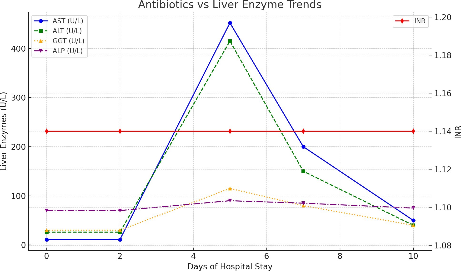
Initial management included levofloxacin for suspected bronchitis, which was later switched to azithromycin after clinical improvement. Atrial fibrillation was treated with a continuous amiodarone drip in the Intensive Care Unit for rate control, along with low dose aspirin due to concerns about ischemic stroke. Anticoagulation therapy with enoxaparin was initially withheld because of the recent cerebrovascular event. On the fifth day of hospitalization, the patient developed increased respiratory distress. Repeat chest imaging revealed new right lung opacities suggestive of an infectious or inflammatory process. Infectious disease specialists recommended starting ceftriaxone 2 g daily intravenously for suspected pneumonia. Shortly after ceftriaxone initiation, serial laboratory testing revealed a dramatic rise in liver enzymes. Baseline liver function tests had been normal (AST 11 U/L, ALT 26 U/L); however, within 48 hours, AST rose to 452 U/L and ALT to 415 U/L. Gamma-glutamyl transferase (GGT) was elevated to 115 U/L, and alkaline phosphatase (ALP) was mildly elevated. PT/PTT and INR remained within normal ranges (PT 12.4, INR 1.14, PTT 30), and hepatitis panel testing was non-reactive. Graphical presentation of the rise and fall of the liver enzymes in relation to antibiotic ceftriaxone is shown in the Figure 1. Total bilirubin was slightly elevated at 0.8 mg/dL. These findings raised concerns for drug induced liver injury (DILI), particularly ceftriaxone induced hepatotoxicity.
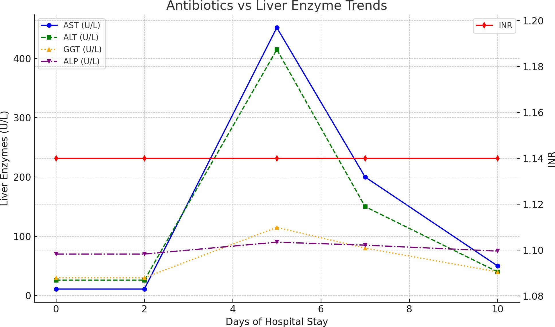
Ceftriaxone was promptly discontinued. Due to the high risk of infection and limited alternative antibiotic options, the patient was transitioned to Aztreonam (1 g intravenously every 8 hours) with continued monitoring of liver enzymes. Daily liver function tests showed a gradual resolution of enzyme elevations following the discontinuation of ceftriaxone. While the patient’s infection improved with the antibiotic switch, her other conditions, including Afib, hypothyroidism, and ischemic stroke, remained stable under routine monitoring and management. This case reinforces the need for vigilance and close monitoring of liver function during ceftriaxone therapy, particularly in vulnerable populations with multiple comorbidities.
DISCUSSION
Mechanism of Action, Metabolism, and Hepatotoxic Potential of Ceftriaxone
Ceftriaxone, a third generation cephalosporin, undergoes minimal hepatic metabolism but is significantly excreted via bile and feces through active transport by hepatocytes.1 Its hepatotoxic potential is influenced by patient related factors, as older age and pre-existing hepatic conditions such as non-alcoholic fatty liver disease (NAFLD) or cholestasis increase susceptibility to liver injury.2 Pharmacodynamically, ceftriaxone exerts its bactericidal activity by binding to penicillin binding proteins (PBPs), thereby inhibiting bacterial cell wall synthesis and leading to cell lysis.3 Elevated biliary concentrations of ceftriaxone can promote the formation of calcium-ceftriaxone complexes, resulting in biliary sludge or obstruction, which contributes to hepatic dysfunction.4 In addition, immune mediated hypersensitivity reactions have been implicated in idiosyncratic cases of ceftriaxone induced liver injury.4 High doses (≥4 g/day) may further exacerbate hepatotoxic risk by saturating biliary excretion pathways, leading to drug accumulation and toxicity.6
This case highlights the potential for ceftriaxone to induce liver injury, even at standard doses, particularly in vulnerable populations such as elderly patients with multiple comorbidities. The rapid elevation of liver enzymes following ceftriaxone administration aligns with findings from other reports. Individual case reports further illustrate the diverse presentations of ceftriaxone induced liver injury, which includes case report by Guarino et al. who reported an elderly patient with acute hepatitis after ceftriaxone administration for pneumonia, with AST and ALT levels peaking at 11,961 U/L and 6,111 U/L, respectively. Cessation of ceftriaxone and supportive care resulted in clinical improvement.5 Nakaharai et al. conducted a retrospective cohort study, revealing that high dose ceftriaxone (≥4 g/day) was associated with a significantly higher incidence of liver injury compared to standard doses (16.2% vs. 2.1%).6 although our patient received a standard dose, her advanced age and comorbidities likely increased her susceptibility to ceftriaxone induced hepatotoxicity. Peker et al. described a 12-year-old boy who developed toxic hepatitis following ceftriaxone therapy, with AST and ALT levels exceeding 800 U/L. Discontinuation of ceftriaxone and corticosteroid administration normalized liver enzymes.7 The clinical course of our patient, marked by significant enzyme elevation and subsequent resolution following ceftriaxone discontinuation, reflects patterns observed in these reports.
Comparison with Liver Failure
Acute liver failure (ALF) is defined by the development of acute liver injury causing hepatic dysfunction, typically with an international normalized ratio (INR) greater than 1.5 and hepatic encephalopathy, in a patient without pre-existing liver disease. In the present case, the patient developed marked elevations in aminotransferases after ceftriaxone exposure, with ALT 415 IU/L and AST 452 IU/L, but these findings alone are insufficient to diagnose ALF. The absence of reported coagulopathy, prolonged PT/INR, jaundice, ascites, or other signs of hepatic decompensation argues against liver failure and is more consistent with isolated drug induced liver injury (DILI). Although hepatic encephalopathy was noted, the overall picture did not meet criteria for acute liver failure because hepatic dysfunction was not accompanied by the full syndrome of ALF, and there was no evidence of multi-organ failure to suggest acute-on-chronic liver failure (ACLF). With discontinuation of ceftriaxone and supportive management, liver enzymes improved, further supporting a reversible DILI pattern rather than progression to liver failure.8–12
This case underscores the multifactorial nature of ceftriaxone induced liver injury. Contributing factors include the patient’s advanced age, reduced hepatic and renal clearance, and possible subclinical cholestasis conditions. These risks, combined with prolonged ceftriaxone therapy, likely triggered the rapid enzyme elevation observed. While the patient’s INR and bilirubin levels remained within normal limits, indicating no immediate progression to acute liver failure, the dramatic rise in AST and ALT levels necessitated discontinuation of ceftriaxone and close monitoring.
CONCLUSION
This case underscores the potential for ceftriaxone induced hepatotoxicity, even at standard doses, particularly in elderly patients with multiple comorbidities. Ceftriaxone’s hepatotoxicity is largely attributed to its biliary elimination and the potential for biliary complications, such as sludge formation and cholestasis, rather than direct metabolic activation. As highlighted by the patient’s clinical course, vigilance is crucial when prescribing ceftriaxone, especially in individuals with predisposing risk factors such as advanced age, polypharmacy, or pre-existing hepatic or renal dysfunction.
The prompt recognition of drug induced liver injury (DILI), immediate discontinuation of ceftriaxone, and transition to an alternative antibiotic (Aztreonam) led to a successful resolution of hepatotoxicity in this case. Regular monitoring of liver function tests during ceftriaxone therapy is essential, particularly in vulnerable populations, to detect and address hepatic complications early. This case also emphasizes the need for clinicians to maintain a high index of suspicion for DILI in patients presenting with elevated liver enzymes during antibiotic therapy.
As ceftriaxone remains a cornerstone of antimicrobial therapy worldwide, balancing its clinical benefits against potential adverse effects requires a multidisciplinary approach. Close collaboration between infectious disease specialists, hepatologists, and pharmacists is critical to optimizing treatment outcomes and minimizing the risk of adverse drug reactions. Furthermore, this case highlights the importance of considering ceftriaxone induced liver injury in patients presenting with elevated liver enzymes during treatment, even at standard doses. Elderly patients with multiple comorbidities may be at increased risk. Regular monitoring of liver function tests during ceftriaxone therapy is essential, and prompt discontinuation should be considered if significant elevations occur. A multidisciplinary approach is vital in managing such cases to balance the benefits of antibiotic therapy against potential adverse effects and this case contributes to the growing body of evidence supporting the importance of regular liver function monitoring and tailored antibiotic selection in high risk patients.
DISCLAIMERS
This article has not been submitted to other publications and/or presented at conferences or meetings.
Source(s) of fund support
None.
Funding Statement
The research received no funding from any source and all authors declare no financial conflict.
Data Availability
The data used in this study was from publicly available published research papers.
Conflict of interest
All authors declare no conflicts of interest.
Regulatory Approval or Research Subject Protection Requirements
Not needed.
Consent form was signed by the patient.
Author contribution
All authors played several overlapping contributory roles such as: Conceptualization, design, cross referencing, and fact checking; Formal Analysis and interpretation of data; project administration, curation, visualization, writing original draft, writing review & editing; supervision, oversight, and leadership, correspondence, data curation, quality control, internal review, communications, data collection and archiving, software, literature search, validation, and approval.